fbpx Skip to main content

Glaucoma Screening

Saint James Eye Clinic offers state-of-the-art facilities to diagnose and treat glaucoma.

What is glaucoma?

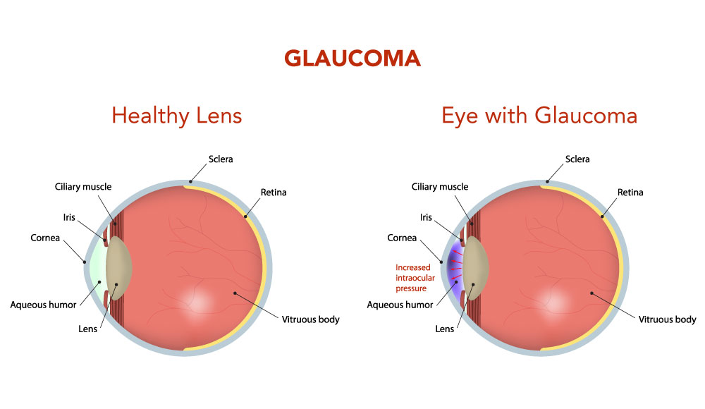
Glaucoma is a condition which can affect sight, usually due to build-up of pressure within the eye. Glaucoma often affects both eyes, usually to varying degrees. One eye may develop glaucoma quicker than the other. The eyeball contains a fluid called aqueous humour which is constantly produced by the eye, with any excess drained through tubes.

Glaucoma develops when the fluid cannot drain properly and pressure builds up, known as the intraocular pressure. This can damage the optic nerve (which connects the eye to the brain) and the nerve fibres from the retina (the light-sensitive nerve tissue that lines the back of the eye).

Glaucoma explained
by Mr Francis Carbonaro

Glaucoma Screening

If you’re at high risk for glaucoma, you should have a dilated pupil eye examination at least every one to two years. To help diagnose glaucoma, an ophthalmologist or optometrist will perform a comprehensive eye exam that may include the following tests.

Tonometry

This test measures the pressure inside the eye. Different types of tonometers include:

  • Air puff or non-contact tonometer — measures the eye’s resistance to a puff of air to determine pressure.
  • Electronic indentation method — measures pressure by directly contacting anesthetized eyes with a digital pen-like instrument.
  • Applanation tonometer — touches the eye’s surface after the eye has been numbed and measures the amount of pressure necessary to flatten the cornea. This is the most sensitive tonometer, but the cornea must be clear and regularly shaped for it to function properly.

Pupil Dilation

Special drops temporarily enlarge the pupil so that the doctor can gain a better view inside of the eye.

Visual Field Testing

This test measures the entire area seen by the forward-looking eye to document straight-ahead (central) and side (peripheral) vision. It measures the dimmest light seen at each spot tested. Each time patients perceive a flash of light, they responds by pressing a button.

Visual Acuity Test

This test measures sight at various distances. The patient reads standardized visual charts with each eye, with and without corrective lenses.

Pachymetry

The eye doctor uses an ultrasonic wave instrument to help determine the thickness of the cornea and better evaluate eye pressure.

Ophthalmoscopy

The doctor examines the interior of the eye by looking through the pupil with a special instrument. This test can help detect damage to the optic nerve caused by glaucoma.

Gonioscopy

The doctor uses this instrument to view the front part of the eye (anterior chamber) to determine if the iris is closer than normal to the back of the cornea. This test can help diagnose closed-angle glaucoma.

Optic Nerve Imaging

Imaging helps document optic nerve changes over time. Nerve imaging can be acquired by Optical Coherence Tomography (OCT).

Types of glaucoma

Chronic open-angle glaucoma

This is the most common type of glaucoma and develops very slowly.

In cases of chronic glaucoma, there are usually no noticeable symptoms because the condition develops very slowly. People don’t often realise their sight is being damaged because the first part of the eye to be affected is the outer field of vision (peripheral vision). Vision is lost from the outer rim of the eye, slowly working inwards towards the centre.

Changes in vision are often linked to getting older, which is why it is so important to have your eyes checked regularly. You should have an eye test at least every two years.

Primary angle-closure glaucoma

This is rare and can occur slowly (chronic) or may develop rapidly (acute) with a sudden, painful build-up of pressure in the eye.

Acute angle-closure glaucoma develops rapidly. Symptoms are often severe and may include:

  • Intense pain
  • Redness of the eye
  • Headache
  • Tender eye area
  • Seeing halos or ‘rainbow-like’ rings around lights
  • Misty vision
  • Loss of vision in one or both eyes that progresses very quickly

As a result of these symptoms, some people may also feel sick or be sick. Symptoms of acute glaucoma are not constant. They can last for one or two hours before disappearing again. But each time the symptoms occur, your vision is damaged a little more. It’s important to seek medical attention if you have any of the above symptoms.

Secondary glaucoma 

This mainly occurs as a result of an eye injury or another eye condition, such as uveitis (inflammation of the middle layer of the eye).

Secondary glaucoma is caused by other conditions, such as uveitis(inflammation of the middle layer of the eye). It can also be caused by eye injuries and certain treatments, such as medication or operations.

It’s possible for the symptoms of glaucoma to be confused with the symptoms of the other condition. For example, uveitis often causes painful eyes and headaches. However, the glaucoma may still cause misty vision and rings or halos around lights.

Developmental glaucoma (congenital glaucoma) 

A rare but sometimes serious type of glaucoma which occurs in very young children, caused by an abnormality of the eye.

Recognising the symptoms of developmental glaucoma (also known as congenital glaucoma) can be difficult due to the young age of the baby or child.

However, your child may display symptoms, such as:

  • Large eyes due to the pressure in the eyes causing them to expand
  • Being sensitive to light (photophobia)
  • Having a cloudy appearance to their eyes
  • Having watery eyes
  • Jerky movements of the eyes
  • Having a squint, which is an eye condition that causes one of the eyes to turn inwards, outwards or upwards, while the other eye looks forward

Preventing glaucoma

Attending regular optician appointments will help to ensure any signs of glaucoma can be detected early and allow treatment to begin.


How common is glaucoma?

Chronic open-angle glaucoma affects up to two in every 100 people over 40 years old and around five in every 100 people over 80 years old. Other types of glaucoma, such as acute angle-closure glaucoma, are much less common.

Risks factors

  • Age. Glaucoma becomes more likely as you get older.
  • Short sightedness (myopia). People who are short-sighted are more likely to develop chronic open-angle glaucoma
  • Ocular hypertension (OHT). Your eye specialist will be able to diagnose OHT which increases your risk of developing chronic open-angle glaucoma.
  • Family history. If you have a close relative, such as a parent, brother or sister who has glaucoma, you are at increased risk of developing the condition yourself
  • Medical history. People with diabetes may be at increased risk of developing glaucoma.

A relatively common cause of secondary glaucoma is known as pseudoexfoliation glaucoma. This type of glaucoma is caused by the body producing abnormal protein fibres, which can block the flow of fluid out of the eye, leading to glaucoma. 

The causes of pseudoexfoliation glaucoma are unclear but most experts think that it is a genetic condition. Pseudoexfoliation glaucoma is treated in the same way as primary glaucoma.

Diagnosing glaucoma

You should have an eye test at least every two years or more frequently if advised by your eye specialist. For example, it may be suggested to you to have more frequent eye tests if you have a close relative with glaucoma.

Tests for glaucoma

There are several glaucoma tests that can be carried out. They are painless and quite quick. The tests should be carried out during the same appointment to ensure results are as accurate as possible.

Eye pressure test (tonometry)

An eye pressure test (tonometry) uses an instrument called a tonometer to measure the pressure inside your eye.

Central corneal thickness

The thickness of your cornea will be measured because this is thought to affect how the intraocular pressure is interpreted.

Gonioscopy

Gonioscopy is an examination of the front outer edge of your eye, between the cornea and the iris (the coloured part of your eye). A gonioscopy can help to determine whether this angle is open or closed (blocked).

Visual field test

A visual field test, sometimes called perimetry, checks for missing areas of vision. You will be shown a sequence of light spots and asked which ones you can see. If you can’t see the spots in your peripheral vision, it may indicate the glaucoma has damaged your vision.

Optical Coherence Tomography (OCT)

The OCT gives accurate information about the retina and the macula. With this information, your eye doctor can recommend the proper treatment, follow the progress of the disease if any, and assess the effectiveness of the treatment.

Optic nerve assessment

Your optic nerve connects your eye to your brain. Your specialist will use eye drops to enlarge your pupils and examine your eyes using a slit lamp to assess whether your optic nerve has been damaged by glaucoma. 

The eye drops used to widen your pupils could affect your ability to drive. You should make alternative arrangements for getting home after your appointment.

Treating glaucoma

Glaucoma can be treated with eye drops, laser treatment or surgery. Early diagnosis is important because damage to the eyes cannot be reversed. 

Treatment aims to reduce the pressure in the affected eye and minimise future damage. Left untreated, glaucoma can cause visual impairment, but if it’s diagnosed and treated early enough, further damage to vision can be prevented.

Eye drops

Chronic open-angle glaucoma is often treated using eye drops. There are several different types of eye drops available.

Laser iridotomy

Laser iridotomy uses a very focused beam of light to create a hole on the outer edge, or rim, of the iris, the coloured part of the eye. This opening may decrease pressure in the eye and usually prevents sudden build-up of pressure within the eye, which occurs during an episode of acute closed-angle glaucoma.

Surgery

A trabeculectomy is the most common type of glaucoma surgery. It involves removing part of the trabecular meshwork to allow fluid to flow through the eye’s drainage system. The procedure will be carried out under local anaesthetic or general anaesthetic.

Other types of surgery include:

  • Viscocanalostomy – this operation removes part of the sclera (the white outer covering of the eyeball), enabling the fluid to filter out of your eye and into your body
  • Deep sclerectomy operation – this operation involves implanting a tiny device inside your eye to widen the trabecular meshwork
  • Aqueous shunt implant – this operation involves placing a tube device into your eye to increase the drainage of fluid out of your eye

Monitoring your condition

If you have been diagnosed with glaucoma your condition will be closely monitored to check for further damage to your vision. Depending on how your glaucoma is progressing, you may need further appointments every one to four months or up to 12-24 months apart.

Complications with glaucoma

The main complication of glaucoma is loss of vision that can’t be repaired. This is why early diagnosis and treatment is so important.

Complications from treatment

If you have surgery to treat glaucoma, there is always a risk of infection. Most infections can be treated with a course of antibiotics. You may also have a reaction to certain types of eye drops.

Other Treatments

Learn More

Glaucoma Screening

A femto laser procedure without a flap to get rid of glasses or contact lenses.

Glaucoma Surgery

A laser procedure to eliminate glasses or contact lenses.

Ready to correct your vision?